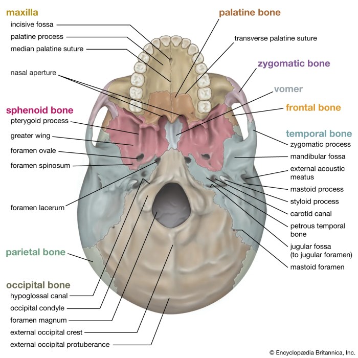
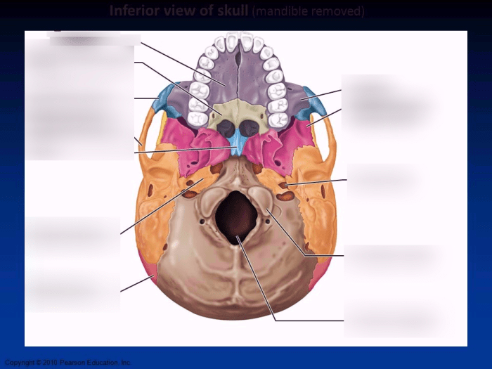
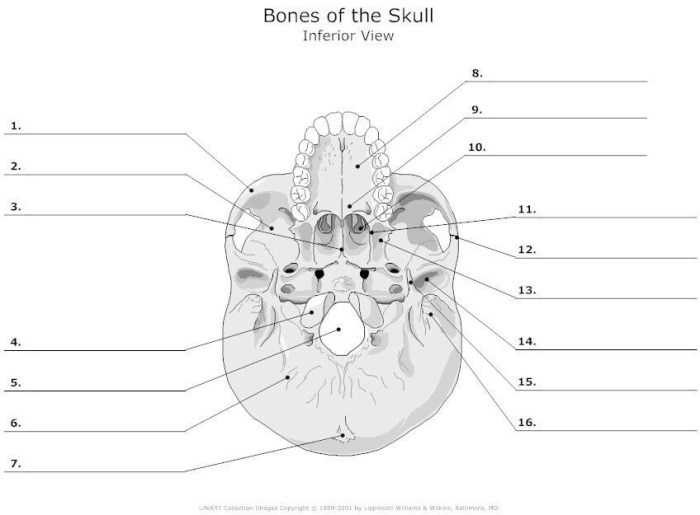
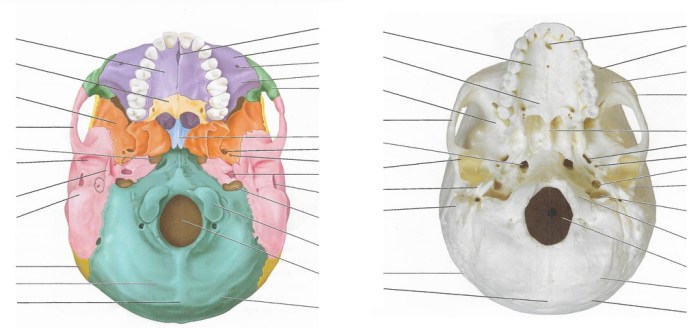
Inferior view of skull mandible removed – The inferior view of the skull with the mandible removed offers a unique perspective of the skull base, revealing intricate anatomical structures and providing valuable insights for clinical practice. This view allows healthcare professionals to examine the skull’s bony landmarks, foramina, muscle attachments, and other features, enabling them to assess cranial nerve function, diagnose skull fractures, and plan surgical interventions.
The inferior view of the skull provides a comprehensive understanding of the skull’s anatomy and its clinical applications. By examining this view, clinicians can gain insights into the complex relationships between different anatomical structures and their functional significance.
Inferior View of Skull: Mandible Removed: Inferior View Of Skull Mandible Removed

The inferior view of the skull, with the mandible removed, provides a comprehensive perspective of the skull’s base. This view allows for the examination of various anatomical structures, including bony landmarks, foramina, muscle attachments, and more.
Bony Landmarks
- Occipital Condyles:Articulate with the first cervical vertebra (atlas) to allow for head movements.
- Foramen Magnum:Large opening in the occipital bone through which the spinal cord passes.
- Mastoid Processes:Projections of the temporal bone behind the ear, containing air cells and the mastoid antrum.
Foramina and Openings
- Jugular Foramen:Located at the base of the temporal bone, transmits the internal jugular vein, glossopharyngeal nerve (CN IX), vagus nerve (CN X), and accessory nerve (CN XI).
- Hypoglossal Canal:Located on the occipital bone, transmits the hypoglossal nerve (CN XII).
- Carotid Canal:Located in the temporal bone, transmits the internal carotid artery.
Muscle Attachments
- Digastric Groove:Groove on the temporal bone for the attachment of the digastric muscle.
- Styloid Process:Projection of the temporal bone for the attachment of muscles, including the stylohyoid, stylopharyngeus, and styloglossus.
- Jugular Process:Projection of the occipital bone for the attachment of the rectus capitis lateralis muscle.
Clinical Applications, Inferior view of skull mandible removed
- Cranial Nerve Assessment:Examination of the foramina on the inferior view of the skull can help assess the function of cranial nerves that pass through them.
- Skull Fracture Diagnosis:Fractures of the skull base can be visualized and assessed from this view.
- Surgical Planning:The inferior view of the skull is essential for planning surgical interventions involving the skull base, such as tumor removal or nerve decompression.
FAQ Corner
What structures are visible on the inferior view of the skull with the mandible removed?
The inferior view of the skull with the mandible removed reveals various anatomical structures, including the occipital condyles, foramen magnum, mastoid processes, jugular foramen, hypoglossal canal, carotid canal, digastric groove, styloid process, and jugular process.
What is the clinical significance of examining the inferior view of the skull?
Examining the inferior view of the skull is clinically significant as it allows healthcare professionals to assess cranial nerve function, diagnose skull fractures, and plan surgical interventions. This view provides insights into the skull’s anatomy and its relationship to surrounding structures, aiding in the diagnosis and management of various clinical conditions.


