Animal Cell Labeled McGraw Hill offers an unparalleled exploration into the intricate world of cells. Embark on a journey of discovery as we delve into the fascinating structures and functions of these fundamental units of life.
Through the comprehensive lens of McGraw-Hill resources, we will unravel the secrets of the cell membrane, cytoplasm, nucleus, and other essential organelles. Prepare to be captivated by the intricate mechanisms that govern cellular processes, shaping the very essence of life.
Introduction
Understanding the structure of animal cells is crucial for comprehending the fundamental processes that govern life. Cells are the basic units of life, and their structure determines their function.
McGraw-Hill resources provide a comprehensive and authoritative foundation for studying cell biology. With clear explanations, engaging visuals, and up-to-date information, McGraw-Hill materials empower students and researchers to delve into the intricate world of cells.
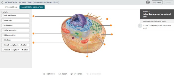
Cell Membrane

The cell membrane, also known as the plasma membrane, is a thin layer that surrounds the cell. It is composed of a phospholipid bilayer, which is a double layer of phospholipids, with the hydrophilic heads facing outward and the hydrophobic tails facing inward.
The cell membrane is selectively permeable, meaning that it allows some substances to pass through while blocking others.
Functions of the Cell Membrane
- The cell membrane regulates the passage of materials into and out of the cell.
- It protects the cell from its surroundings.
- It helps the cell maintain its shape.
- It is involved in cell signaling.
Cytoplasm

The cytoplasm is the jelly-like substance that fills the cell and surrounds the nucleus. It is composed of about 70% water, and the rest is a complex mixture of proteins, carbohydrates, lipids, and other molecules. The cytoplasm is the site of many important cellular activities, including protein synthesis, cell division, and energy production.
Organization of the Cytoplasm
The cytoplasm is not a uniform substance, but rather is organized into a number of different compartments. These compartments include the endoplasmic reticulum, the Golgi apparatus, the mitochondria, and the lysosomes. Each of these compartments has a specific function, and they work together to maintain the cell’s homeostasis.
Role of the Cytoplasm in Cellular Activities
The cytoplasm plays a vital role in many cellular activities. It is the site of protein synthesis, which is essential for cell growth and repair. The cytoplasm also contains the mitochondria, which are the cell’s energy producers. In addition, the cytoplasm is involved in cell division and cell movement.
Nucleus
The nucleus, often referred to as the control center of the cell, is a membrane-bound organelle found in eukaryotic cells. It plays a crucial role in directing and regulating cellular activities.
The nucleus is enclosed within a double-layered membrane called the nuclear envelope, which separates it from the cytoplasm. Within the nucleus, we find the genetic material, DNA, organized into structures called chromosomes. These chromosomes contain genes, which are the units of heredity that determine the characteristics of an organism.
Functions of the Nucleus
The nucleus is responsible for a wide range of functions that are essential for cellular life, including:
- Gene Expression:The nucleus controls the expression of genes by regulating which genes are transcribed into messenger RNA (mRNA). mRNA carries the genetic information from the nucleus to the cytoplasm, where it is translated into proteins.
- DNA Replication:During cell division, the nucleus ensures that each new cell receives an identical copy of the genetic material. This process is called DNA replication and occurs before cell division.
- Cellular Regulation:The nucleus plays a key role in regulating cellular activities by producing proteins that function as enzymes, hormones, and structural components.
- Ribosome Production:The nucleus contains structures called nucleoli, which are responsible for producing ribosomes. Ribosomes are the cellular machinery that synthesizes proteins.
Mitochondria
Mitochondria are small organelles found in the cytoplasm of eukaryotic cells. They are often referred to as the “powerhouses of the cell” because they are responsible for generating most of the cell’s energy. Mitochondria have a double membrane structure, with the inner membrane folded into cristae.
These cristae provide a large surface area for the enzymes involved in cellular respiration to attach to.
Function of Mitochondria, Animal cell labeled mcgraw hill
The main function of mitochondria is to produce adenosine triphosphate (ATP), the cell’s main energy currency. ATP is used to power all of the cell’s activities, from muscle contraction to protein synthesis. Mitochondria produce ATP through a process called oxidative phosphorylation, which involves the transfer of electrons from NADH and FADH2 to oxygen.
This process creates a proton gradient across the inner mitochondrial membrane, which is used to drive the synthesis of ATP.Mitochondria also play a role in other cellular processes, such as calcium buffering, apoptosis (programmed cell death), and the synthesis of heme.
Endoplasmic Reticulum
The endoplasmic reticulum (ER) is an extensive network of membranes that folds and transports proteins. It is composed of two types: rough ER and smooth ER. The rough ER is studded with ribosomes, which are responsible for protein synthesis. The smooth ER is involved in lipid metabolism and detoxification.
Exploring the intricacies of animal cell structures through McGraw Hill’s labeled diagrams offers a comprehensive understanding of cellular components. To further enhance your culinary knowledge, we recommend visiting the parts and labor butchery menu for a detailed exploration of animal anatomy and its culinary applications.
Returning to the topic of animal cell labeling, McGraw Hill’s resources provide invaluable insights into the fundamental units of life.
Structure of the Endoplasmic Reticulum
The ER is a continuous membrane system that extends throughout the cytoplasm. It is composed of a network of flattened sacs called cisternae. The cisternae are surrounded by a single membrane that is continuous with the nuclear envelope. The ER is also connected to the Golgi apparatus by a series of vesicles.
Functions of the Endoplasmic Reticulum
The ER has a variety of functions, including:* Protein synthesis:The rough ER is the site of protein synthesis. Ribosomes attach to the ER membrane and translate mRNA into proteins. The proteins are then folded and transported to the Golgi apparatus.
Lipid metabolism
The smooth ER is involved in lipid metabolism. It synthesizes lipids and steroids and also detoxifies drugs and other harmful substances.
Golgi Apparatus

The Golgi apparatus, also known as the Golgi complex or Golgi body, is an essential organelle found in eukaryotic cells. It is a stack of flattened, membrane-bound sacs called cisternae. The Golgi apparatus plays a crucial role in modifying, sorting, and packaging proteins synthesized in the cell.
Protein Modification
As proteins exit the rough endoplasmic reticulum (RER), they enter the Golgi apparatus. Within the Golgi cisternae, proteins undergo various modifications, including:
- Glycosylation: Addition of sugar molecules to proteins.
- Phosphorylation: Addition of phosphate groups to proteins.
- Sulfation: Addition of sulfate groups to proteins.
These modifications alter the protein’s structure and function, preparing them for their specific roles within the cell.
Protein Sorting
The Golgi apparatus also functions as a sorting and packaging center for proteins. Proteins are sorted based on their destination and enclosed in membrane-bound vesicles. These vesicles then bud off from the Golgi and are transported to their target locations, such as the plasma membrane, lysosomes, or secretory vesicles.
Lysosomes
Lysosomes are membrane-bound organelles found in the cytoplasm of animal cells. They are small, spherical vesicles that contain a variety of hydrolytic enzymes, which are capable of breaking down a wide range of biomolecules, including proteins, carbohydrates, lipids, and nucleic acids.
Lysosomes play a crucial role in cellular digestion. They engulf and fuse with endocytic vesicles, which contain materials taken up by the cell from the external environment. The hydrolytic enzymes within the lysosomes then break down the contents of these vesicles, releasing the nutrients that the cell can use for energy or to build new molecules.
Role in Cellular Digestion
Lysosomes are essential for the proper functioning of cells. They help to remove waste products and damaged organelles, and they play a role in the recycling of cellular components. Lysosomal dysfunction has been linked to a number of diseases, including lysosomal storage diseases, which are characterized by the accumulation of undigested materials within lysosomes.
Ribosomes
Ribosomes are tiny, complex organelles found in all living cells. They are responsible for protein synthesis, one of the most essential processes in cell biology.Ribosomes are composed of two subunits: a large subunit and a small subunit. Each subunit is made up of a combination of ribosomal RNA (rRNA) and proteins.
The large subunit contains the catalytic center where protein synthesis occurs.
Role of Ribosomes in Protein Synthesis
Ribosomes play a crucial role in protein synthesis by assembling amino acids into polypeptide chains. This process is divided into three main steps: initiation, elongation, and termination.
- Initiation:The ribosome binds to a messenger RNA (mRNA) molecule, which carries the genetic code for the protein to be synthesized. The small subunit binds to the mRNA and locates the start codon (AUG), which signals the beginning of protein synthesis.
- Elongation:The ribosome moves along the mRNA molecule, reading the codons and adding the corresponding amino acids to the growing polypeptide chain. Transfer RNA (tRNA) molecules carry the amino acids to the ribosome and align them with the correct codons on the mRNA.
- Termination:When the ribosome reaches a stop codon on the mRNA, it releases the completed polypeptide chain and dissociates into its two subunits.
Ribosomes are essential for cell function and survival. They are responsible for synthesizing all the proteins that the cell needs to function properly, including enzymes, hormones, and structural proteins. Without ribosomes, cells would not be able to produce the proteins necessary for life.
Cytoskeleton: Animal Cell Labeled Mcgraw Hill
The cytoskeleton is a dynamic network of protein filaments and tubules that extends throughout the cytoplasm of eukaryotic cells. It provides structural support, maintains cell shape, and facilitates cellular movement.The cytoskeleton is composed of three main types of filaments: microtubules, microfilaments, and intermediate filaments.
Microtubules are long, hollow cylinders made of tubulin protein subunits. They are responsible for maintaining cell shape and providing tracks for organelle movement. Microfilaments are thin, solid rods made of actin protein subunits. They are involved in cell movement, such as muscle contraction and cell crawling.
Intermediate filaments are thicker and more flexible than microtubules and microfilaments. They help to maintain cell shape and resist mechanical stress.The cytoskeleton is a highly dynamic structure that constantly changes in response to cellular needs. It is essential for many cellular processes, including cell division, cell movement, and cell signaling.
Expert Answers
What is the significance of studying animal cell structure?
Understanding animal cell structure is crucial for comprehending the fundamental processes of life. Cells are the basic building blocks of all living organisms, and their structure and function determine the overall health and functioning of the organism.
How do McGraw-Hill resources contribute to the study of cell biology?
McGraw-Hill resources provide a wealth of high-quality educational materials, including textbooks, online resources, and interactive simulations. These resources are designed to support students and researchers in their exploration of cell biology, making complex concepts accessible and engaging.